Forward Scatter vs Side Scatter
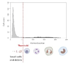
Forward Threshold is very important in order to eliminate from the pool data, the particules that should be ignored from the data analisys.
Whenever a particle or cell passes through the laser beam a voltage pulse is generated.
To prevent interference from background noise or debris, a threshold can be set.
By setting a threshold signal value, processing only occurs when a voltage pulse signal is above this limit. Signals below threshold are not processed.
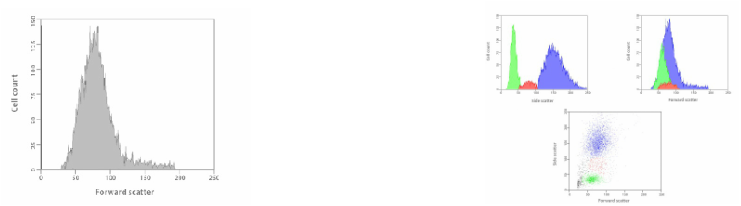
By gating only the Forward Scatter ( FSC) we will have a one dimensional histogram were can be only be seen at the range size of the particules in the sample.
On the other hand gating the Side Scatter (SSC) with the FSC we can get an upgrade to a two dimensional histograms or scatter dot plots.
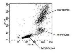
For example, using the FSC and the SSC we can obtain the division of the blood cells arranged by gates of complexity and size.The sample was a lysed whole blood.
The forward and side scatter light signals are emitted at a 488 nm wavelength and are of the same colour as the laser light. These signals can therefore be determined without the need to stain with dyes or fluorochromes.
However for a better and complete use of this multiparametric analysis is necessary to apply Flurescence technics.
Fig. http://probes.invitrogen.com/resources/education/tutorials/4Intro_Flow/player.html
Reference:Leach, M., Drummond, M., and Doig, A. (2013) Pratical Flow Cytometry in Haematology Diagnosis. John Wiley & Sons, Ltd.
http://www.stemcell.umn.edu/prod/groups/med/@pub/@med/documents/asset/med_80691.pdf
